
Total Items
0
0 Item(s)

Blog post
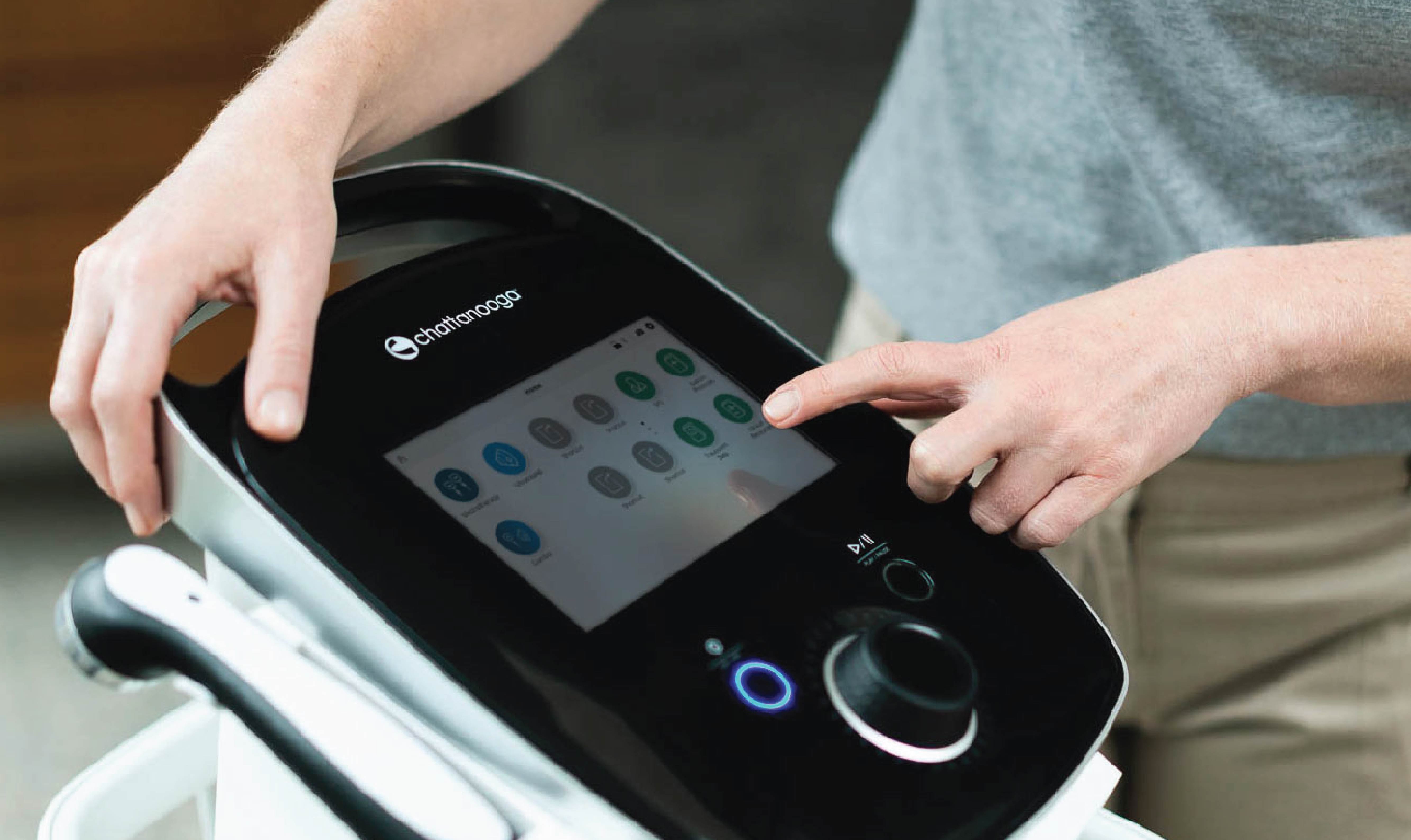
Meet the Next Generation of ElectrotherapyIntroducing the Chattanooga® Intelect® Legend 2 & Intelect® Transport 2 In February, Enovis™ announced the launch of the Chattanooga® Intelect® Legend 2 and Intelect® Transport 2 devices. These two products build on the legacy of Chattanooga electrotherapy products known for their durability and reliability as staples in the sports medicine and rehabilitation community for over 70 years. Dating back to the origins of the Chattanooga brand in 1947, Enovis has focused the design of the Intelect Legend 2 and the Intelect Transport 2 around two basic tenets – helping patients not only recover from injury but return to activities of daily living in less time and allowing clinicians to devote more time to their patients. Image The Intelect Legend 2 and Intelect Transport 2 are powered by Variable Muscle Stimulation (VMS) – a proprietary waveform exclusive to Chattanooga electrotherapy products. VMS is a symmetrical biphasic waveform offering several advantages over other waveforms when treating patients with neuromuscular electrical stimulation. VMS has been shown to elicit greater muscle contractions without impacting patient comfort when compared to the commonly used Russian waveform.1 VMS has also been shown to produce less muscle fatigue which can lead to more effective strength training sessions.2 For more information on VMS, check out our latest webinar VMS for NMES: Uncovering Advantages Over Russian Waveform, or our new VMS White Paper, courtesy of Enovis key opinion leader James Bellew, PT, EdD, MS.* In addition to significant evidence supporting the benefits of VMS,1,2 the Intelect Legend 2 provides clinicians with the ability to set up treatment in just three button pushes with its easy-to-use Suggested Protocol Settings (SPS) menu to reduce patient setup time. The Intelect Legend 2’s built-in SPS menu allows clinicians to confidently set up appropriate treatments based on suggested, evidence-based protocols for the indication and the patient’s condition. To read Enovis’s full press release on the commercial launch of the Intelect Legend 2 and Intelect Transport 2, please visit https://enovis.com/investors/press-releases/enovis-showcase-new-intelectr-legend-2-and-intelectr-transport-2. *James Bellew, PT, EdD, MS is a consultant for Enovis. The opinions and experiences presented here are for informational purposes only. James Bellew, PT, EdD, MS has been compensated by Enovis for time and effort expended in preparing and presenting this webinar for Enovis’ further use and distribution. References Bellew JW, Allen M, Biefnes A, Grantham S, Miglin J, Swartzell D. Efficiency of neuromuscular electrical stimulation: A comparison of elicited force and subject tolerance using three electrical waveforms. Physiotherapy Theory and Practice. 2018;34(7):551-558. doi:10.1080/09593985.2017.1422820 Cayot TE, Bellew JW, Kennedy S, Pursley E, Smith N, Stemme K. Acute effects of 3 neuromuscular electrical stimulation waveforms on exercising and recovery microvascular oxygenation responses. Journal of Sport Rehabilitation. 2022;31(5):554-561. doi:10.1123/jsr.2021-0326
Blog post
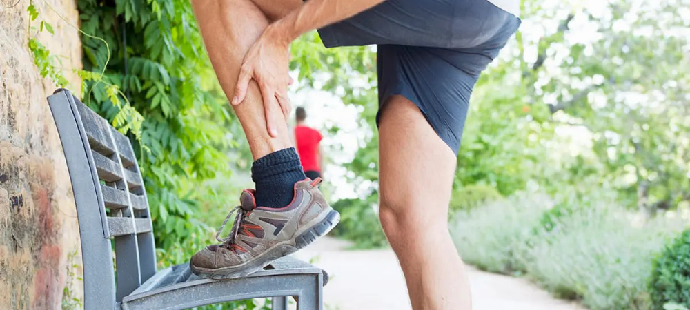
Introduction of Extracorporeal Shockwave Therapy (ESWT) for Running InjuriesImage Soft tissue injuries are common to runners and athletes who depend on their legs to get them where they need to go. About 65% of regular runners get hurt each year. It’s estimated that the average runner will sustain one injury for every 100 hours he or she runs.1 When an injury occurs, athletes and weekend warriors are usually looking for solutions to get them back on the road as soon as possible. What is shockwave? They are high-energy waves created by sharp changes of pressure in a narrow region, traveling through a medium like air or water. They are normally caused by an explosion or by a body moving faster than sound.2 How does shockwave help tissue? High energy waves, that can be created via different mechanisms, create a phenomenon referred to as mechanotransduction. In simple terms, it is the process of imparting brief, physical deformation to cells that lead to biochemical changes. These changes have the potential to positively impact pain and tissue repair.3 In some instances, the negative pressure created during the tensile phase of a shockwave creates cavitation bubbles within cells.4 If intense enough, it can lead to disruption of damaged cells which is why shockwave can be classified as a proinflammatory modality. Disruption of cells can lead to cell death (apoptosis) which triggers a low-level inflammatory response that benefits the process of removal and replacement of damaged tissue. This is another way that shockwave therapy can uniquely assist in treating chronic soft tissue problems. Terminology Names that refer to therapeutic shockwave are varied. This can create confusion when trying to investigate this technology. Common names include but are not limited to: ESWT: Extracorporeal Shockwave Therapy FSW: Focused Shockwave EPAT: Extracorporeal Pulsed Activation Therapy RPW: Radial Pressure Wave AWT: Acoustic Wave Therapy The list seems to grow by the month as companies try to differentiate their products. Classification of FSW vs RPW Generally speaking, there are two primary families of devices under the ESWT umbrella, the focused shockwave (FSW) and radial pressure wave (RPW) devices. While the two waves possess different physical characteristics, they generate similar results when treating various conditions in the musculoskeletal system with equivalent dosing.5, 6 Definition of shockwave components and characteristics A FSW is a high-intensity, low-frequency (1 to 8 Hz) wave that impacts tissue differently than therapeutic ultrasound (US). Therapeutic shockwaves are non-thermal waves that create mechanotransduction and in some cases cavitation in tissues as deep as 12 cm.4, 7 FSW energy delivery is measured in mJ/mm2 which is referred to as Energy Flux Density (EFD). EFD ranges between 0.01 and 0.55 mJ/mm2 on Chattanooga FSW equipment.8 Preferable dosing ranges exist when treating different conditions. For example, treating with extremely low energy levels (below 0.08 mJ/mm2) has been shown to be ineffective and/or less effective than treating in higher energy ranges.6, 9 On the other end of the spectrum, treating at energy levels >0.60 mJ/mm2 has been shown to be deleterious to tendons.9 Understanding correct dosing parameters and treatment approaches is imperative to achieving consistent results with this equipment. Interpreting bar pressure vs energy flux density (EFD) RPW devices are commonly measured in bar pressure. This is likely due to the pneumatic mechanism that generates pressure waves. Bar pressure should ideally be measured at the point where the applicator meets the skin to ensure accurate clinical relevance. 1 bar is equivalent to 14.5 psi. With the correct conversion factors, bar pressure can be converted to EFD. This allows for equivalent dosing parameters to be calculated when comparing RPW and FSW treatments of a given condition. An example of a radial pressure wave is pictured below next to a traditional focused shockwave for ease of comparison. Notice the differences in peak pressure, rate of the wave cycle, as well as the general shape of the 2 waves. Image Image Fig 1. What are shock waves? Physics and Technology. STORZ MEDICAL - The Shock Wave Company. Accessed January 9, 2024. https://www.storzmedical.com/us/physics-and-technology The physical impact of shockwaves can help improve the environment surrounding recalcitrant problems in muscle, calcific tendons, plantar fasciitis, as well as different connective tissues. Are all soundwaves the same? Some clinicians falsely assume that shockwave equipment is similar to therapeutic ultrasound (US) since they both utilize soundwaves. It should be noted that therapeutic ultrasound uses a lower intensity sound wave (20 to 1000 mW/cm2) that is delivered at a higher frequency (0.7 to 3.3 MHz.).10 Therapeutic US waves look like this: Image Fig 2. What are shock waves? Physics and Technology. STORZ MEDICAL - The Shock Wave Company. Accessed January 9, 2024. https://www.storzmedical.com/us/physics-and-technology Continuous US creates thermal effects in tissue by alternating compression and rarefaction of sound waves within tissue. Maximum energy absorption in soft tissue occurs from 2 to 5 cm and intensity decreases as the waves penetrate deeper.10 Hopefully this review will provide some clarity for readers that are looking to better understand how ESWT can help various conditions as well as which device is best suited for a given practice setting. References Running Injuries. Yale Medicine. https://www.yalemedicine.org/conditions/running-injury#:~:text=About%2065%20percent%20of%20regular%20runners%20get%20hurt shockwave definition. Bing. Accessed December 18, 2023. https://www.bing.com/search?q=shockwave+definition&cvid=fc98a1ef85b74e16acacd38d178a34da&gs_lcrp=EgZjaHJvbWUqBggAEAAYQDIGCAAQABhAMgYIARBFGDkyBggCEAAYQDIGCAMQABhAMgYIBBAAGEAyBggFEAAYQDIGCAYQABhAMgYIBxAAGEAyBggIEAAYQDIICAkQ6QcY8gcyBwgKEEUY_FXSAQgyMTY5ajBqNKgCALACAA&FORM=ANAB01&PC=U531 d’Agostino MC, Craig K, Tibalt E, Respizzi S. Shock wave as biological therapeutic tool: From mechanical stimulation to recovery and healing, through mechanotransduction. Int J Surg. 2015;24(Pt B):147-153. doi:10.1016/j.ijsu.2015.11.030 Fuchs J. Stable vs. Transient Cavitation. CTG Technical Blog. Published January 17, 2019. Accessed December 18, 2023. https://techblog.ctgclean.com/2019/01/stable-vs-transientcavitation/#:~:text=Basically%2C%20the%20cavity%20is%20created%20but%20the%20contents Schroeder AN, Tenforde AS, Jelsing EJ. Extracorporeal Shockwave Therapy in the Management of Sports Medicine Injuries. Current Sports Medicine Reports. 2021;20(6):298-305. doi:https://doi.org/10.1249/JSR.0000000000000851 Schmitz C, Császár NB, Milz S, et al. Efficacy and safety of extracorporeal shock wave therapy for orthopedic conditions: a systematic review on studies listed in the PEDro database. Br Med Bull. 2015;116(1):115-138. doi:10.1093/bmb/ldv047 RPW User Manual. https://enovis-medtech.eu/media/storage.djoglobal.eu/en_US/Documents/Support_documents/IFU_13-28670_US_Rev_A_Intelect_RPW_2_DIGITAL_Final.pdf Focus Shock Wave User Manual. https://www.djoglobal.eu/media/storage.djoglobal.eu/en_US/Documents/Documents_2023/13-00061-US_RevE_USA_IFU,_FOCUS_SHOCKWAVE-EN.pdf Rompe JD, Kirkpatrick CJ, Küllmer K, Schwitalle M, Krischek O. Dose-related effects of shock waves on rabbit tendo Achillis. A sonographic and histological study. J Bone Joint Surg Br. 1998;80(3):546-552. doi:10.1302/0301-620x.80b3.8434 Uddin SMZ, Komatsu DE, Motyka T, Petterson S. Low-Intensity Continuous Ultrasound Therapies—A Systematic Review of Current State-of-the-Art and Future Perspectives. J Clin Med. 2021;10(12):2698. Published 2021 Jun 18. doi:10.3390/jcm10122698
Blog post
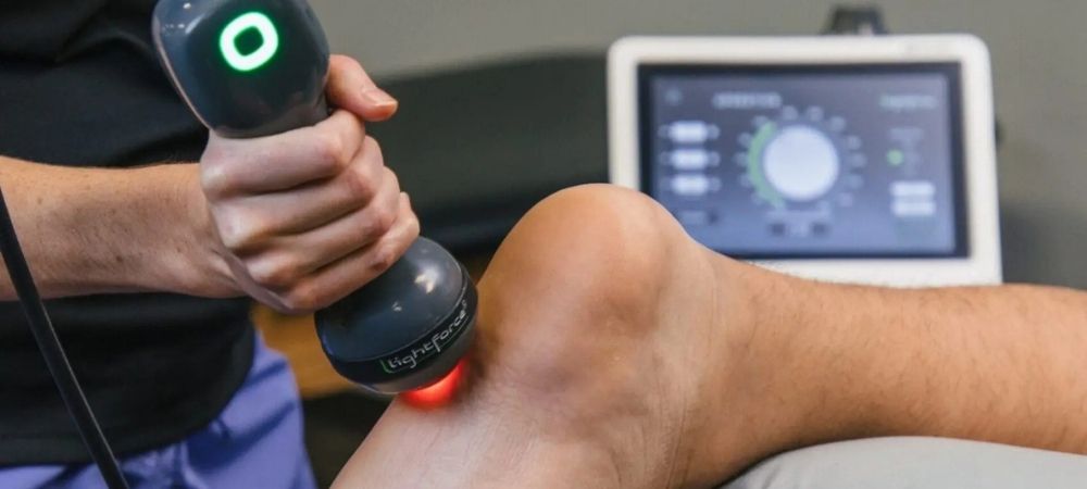
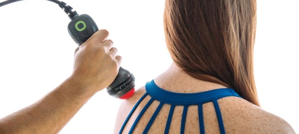
Revised 2023 APTA Clinical Practice Guidelines for Heel Pain – Plantar Fasciitis Upgrades Therapeutic Laser RecommendationClinicians should use laser therapy to decrease pain for acute and chronic plantar fasciitis Laser therapy received a grade B recommendation in the newly released 2023 American Physical Therapy Association (APTA) clinical practice guidelines (CPG) for managing plantar fasciitis. This is an upgrade from a grade C recommendation in the 2014 guidelines.1 During the past 10 years, a substantial amount of peer-reviewed literature has been published supporting laser therapy as a treatment option for musculoskeletal pain. We are pleased to see that APTA recognizes the quality scientific evidence published over the last decade and has determined to give laser therapy a higher recommendation in its latest CPG. The upgraded rating means that physical therapists should no longer just consider adding laser therapy to their treatment protocol for acute and chronic plantar fasciitis, but, in fact, should be offering this treatment modality to help improve their patient’s pain.1 Click here to access the CPG. Image Ready to add LightForce® laser therapy to your clinic? Laser therapy can be used for more than just plantar fasciitis. LightForce® lasers are FDA cleared to treat minor muscle and joint pain, joint stiffness, arthritis pain, and muscle spasms so there is a multitude of ways that you can incorporate laser therapy to help give your patients a better outcome. In addition to plantar fasciitis, the APTA recognizes laser therapy as a treatment option for acute ankle pain and acute and chronic neck pain.2,3 The American College of Physicians includes laser therapy in its recommendations for chronic low back pain.4 Laser therapy is a modality backed by scientific research and supported by clinical evidence to relieve musculoskeletal pain. To speak to a Chattanooga® representative or to request a demo, please visit https://www.chattanoogarehab.com/us/laser-therapy/. References Koc TA Jr, Bise CG, Neville C, Carreira D, Martin RL, McDonough CM. Heel Pain – Plantar Fasciitis: Revision 2023. J Orthop Sports Phys Ther. 2023;53(12):CPG1-CPG39. doi:10.2519/jospt.2023.0303 Martin RL, Davenport TE, Fraser JJ, et al. Ankle Stability and Movement Coordination Impairments: Lateral Ankle Ligament Sprains Revision 2021. J Orthop Sports Phys Ther. 2021;51(4):CPG1-CPG80. doi:10.2519/jospt.2021.0302 Blanpied PR, Gross AR, Elliott JM, et al. Neck Pain: Revision 2017. J Orthop Sports Phys Ther. 2017;47(7):A1-A83. doi:10.2519/jospt.2017.0302 Qaseem A, Wilt TJ, McLean RM, et al. Noninvasive Treatments for Acute, Subacute, and Chronic Low Back Pain: A Clinical Practice Guideline From the American College of Physicians. Ann Intern Med. 2017;166(7):514-530. doi:10.7326/M16-2367 Read More Blog Posts
Blog post
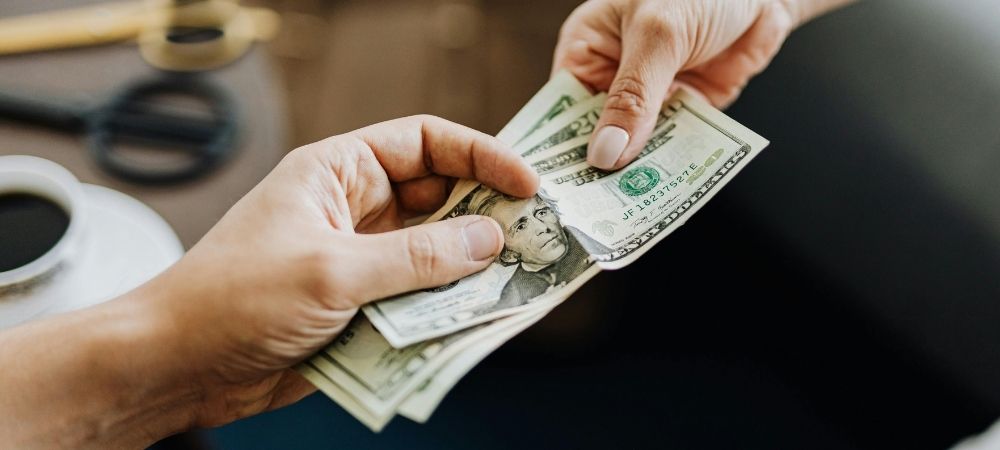
3 Cash-Based Services for Private PracticesContributed by Mark Callanen, PT, DPT, OCS Cash-based services should add unique and discernible value to your patients' plan of care. If your patients feel a difference within just a few treatments, the value of this service will exceed its costs. For this reason, selecting cash services that focus on quickly reducing pain complaints is a smart strategy. Chattanooga® offers numerous medical devices that help decrease pain and improve patient outcomes. Three popular cash treatment options are in the areas of therapeutic laser, radial pressure wave therapy, and traction/ decompression treatments. While each device imparts different mechanisms to help treat various conditions, they all share the ability to reduce pain in short periods of time.1-3 LightForce® Therapy Lasers LightForce® Therapy Lasers are one of the most popular devices used in the cash pay market. Key reasons include: Laser can be applied to chronic and acute conditions and is well tolerated by most patients.4 It has the ability to reduce minor muscle and joint pain, muscle spasm, as well as pain and stiffness associated with arthritis. Laser assists with tissue repair by promoting relaxation of muscle tissue and temporarily increasing local blood circulation. Treatment can be applied as cash supplement to most treatment sessions as the billing codes associated with laser (LLLT) are generally not recognized by third party payers. **(Check with billing professional) Chattanooga® Radial Pressure Wave Therapy Radial Pressure Wave therapy is another non-invasive treatment option that uses high intensity sound waves to impact pain and increase blood flow to various tissues. It has been shown to help treat a variety of soft tissue dysfunctions. These include, but are not limited to, trapezius muscle pain, shoulder pain, elbow pain, knee pain, Achilles tendon pain, or plantar and heel pain. Primary benefits include: Radial pressure waves have the ability to effectively reach pain generating tissues at up to 6 cm. Large areas can be scanned and treated in a short treatment time. Most plans of care consist of about 3 treatments.5 Treatment tool for locating and treating active and latent trigger points.2 Pain can be impacted after a single treatment.2 Treatment can be applied as cash supplement to most treatment sessions as the billing codes associated with shockwave treatments (ESWT) are generally not recognized by third party payers. **(Check with billing professional) Chattanooga® Traction/ Decompression Therapy Low back pain (LBP) is one of the most common reasons adults in the US seek out a primary care doctor. A 2020 study showed that 55% of LBP patients present with intervertebral narrowing of the lumbar spine. Additionally, 25% of LBP patients present with radicular complaints into the lower legs.6 Intervertebral narrowing and radicular complaints are both associated with disc pathology. Traction has been shown to decrease disc bulging, improve intervertebral narrowing,7 and to be a more effective treatment option for reducing pain and disability scores than exercise alone for patients with disc pathology.8 Chattanooga® traction devices are a potential treatment option for these patients. A 2023 post market survey of 272 low back pain cases found that Chattanooga® traction devices were 87.5% effective in reducing low back pain.9 Primary benefits of incorporating traction therapy include: Can be used for chronic LBP with radicular complaints.8 Can provide predictable amounts of tension to help decompress spinal structures.7,10 Treatments are easily replicated. Can provide longer duration and higher force traction that is difficult to perform manually. Is an unattended treatment that helps reduce pain.9 Ability to charge cash for this service is dependent on reimbursement rules for various carriers. ** (Check with your billing professional) All three of these solutions address different types of patient conditions. However, they are all focused at reducing pain and providing treatment options for patients looking to avoid surgery and/ or reduce the need to rely on pain medications. Many practice owners in the Breakthrough community have adopted one or more of these technologies with great results. Practice owner Tony Cere has added $300,000 in annual cash-based revenue. “Patients will come in for the laser, and turn into physical therapy patients,” he said. According to Tom Loyd, Practice Owner at Bryn Mawr Sports Therapy, “The Lightforce 40-watt laser generates improved patient outcomes and adds an accessory form of income.” References Fritz JM, Lindsay W, Matheson JW, et al. Is there a subgroup of patients with low back pain likely to benefit from mechanical traction? Results of a randomized clinical trial and subgrouping analysis. Spine (Phila Pa 1976). 2007;32(26):E793-E800. doi:10.1097/BRS.0b013e31815d001a Kartaloglu IF, Kus AA. Evaluation of Radial Extracorporeal Shock Wave Therapy on Treatment-Resistant Trigger Points Using Sonographic Shear Wave Elastography. J Coll Physicians Surg Pak. 2023;33(10):1159-1164. doi:10.29271/jcpsp.2023.10.1159 Chow RT, Johnson MI, Lopes-Martins RA, Bjordal JM. Efficacy of low-level laser therapy in the management of neck pain: a systematic review and meta-analysis of randomised placebo or active-treatment controlled trials [published correction appears in Lancet. 2010 Mar 13;375(9718):894]. Lancet. 2009;374(9705):1897-1908. doi:10.1016/S0140-6736(09)61522-1 Arroyo-Fernández R, Aceituno-Gómez J, Serrano-Muñoz D, Avendaño-Coy J. High-Intensity Laser Therapy for Musculoskeletal Disorders: A Systematic Review and Meta-Analysis of Randomized Clinical Trials. J Clin Med. 2023;12(4):1479. Published 2023 Feb 13. doi:10.3390/jcm12041479 Schmitz C, Császár NB, Milz S, et al. Efficacy and safety of extracorporeal shock wave therapy for orthopedic conditions: a systematic review on studies listed in the PEDro database. Br Med Bull. 2015;116(1):115-138. doi:10.1093/bmb/ldv047 Kamal KC, Alexandru DO, Kamal D, et al. Managing Low Back Pain in Primary Care. Curr Health Sci J. 2020;46(4):396-404. doi:10.12865/CHSJ.46.04.11 Chung TS, Yang HE, Ahn SJ, Park JH. Herniated Lumbar Disks: Real-time MR Imaging Evaluation during Continuous Traction [published correction appears in Radiology. 2015 Jun;275(3):934-5]. Radiology. 2015;275(3):755-762. doi:10.1148/radiol.14141400 Wang W, Long F, Wu X, Li S, Lin J. Clinical Efficacy of Mechanical Traction as Physical Therapy for Lumbar Disc Herniation: A Meta-Analysis. Comput Math Methods Med. 2022;2022:5670303. Published 2022 Jun 21. doi:10.1155/2022/5670303 (2023). Post-Market Clinical Follow-Up Retrospective Study Report: Traction and Decompression Therapy Systems. Internal Enovis report. Unpublished. Chow DHK, Yuen EMK, Xiao L, Leung MCP. Mechanical effects of traction on lumbar intervertebral discs: A magnetic resonance imaging study. Musculoskelet Sci Pract. 2017;29:78-83. doi:10.1016/j.msksp.2017.03.007 Read More Blog Posts
Blog post
DJO AnnouncementDJO® Acquires LiteCure Laser Therapy Acquisition establishes Chattanooga® as the leader in high-power laser therapy Dallas, TX (December 11, 2020) – DJO, LLC (“DJO” or the “Company”), a leading global provider of medical technologies to get and keep people moving, today announced the acquisition of LiteCure®, the market leader in therapeutic laser technology for human and animal health. This purchase further extends DJO’s strength as the global market leader in recovery sciences and allows the company to deliver unparalleled innovation to its customers and their patients. “The LiteCure portfolio was a natural fit for the DJO Recovery Sciences business, bringing together a joint passion for innovation and clinically proven technology,” said Brady Shirley, DJO CEO. “The acquisition will strengthen our leadership position in physical therapy and rehabilitation – and reinforces our commitment to keeping people active; enabling our customers to better serve their patients.” DJO has long respected LiteCure’s reputation as the market leader in laser light therapy and identified immediate synergies with its Chattanooga rehabilitation business. “The LiteCure portfolio complements and expands Chattanooga’s innovative rehabilitation therapies,” said Terry Ross, President, DJO Recovery Sciences. “Together, we will bring leading technologies and treatment modalities designed to enhance patient outcomes and practice efficiency to our customers around the world.” LiteCure’s products are marketed under the brand names of LightForce® Therapy Lasers for humans and Companion® for animals, which includes Pegasus® equine laser technology. LightForce is the industry leader in medical therapy laser manufacturing and innovation, and continually seeks to move the industry forward through laser science, solutions and technology. LightForce Therapy Lasers can be found in over 250 professional and college athletic training rooms and are the preferred modality when treating world-class athletes. With positive clinical results, high power lasers are one of the fastest growing modalities in physical therapy. In addition, the Companion brand provides a strong entry point into the thriving animal health space and brings with it an accomplished, dedicated animal health team. DJO plans to leverage parts of its existing product portfolio to create an immediate impact in animal health while working with the team to explore incremental growth opportunities. “With DJO’s expanded reach and resources, we are excited to increase awareness and adoption of deep tissue laser therapy,” added Brian Pryor, Founder and CEO of LiteCure. “We are passionate about the clinical benefits of photobiomodulation as well as the benefits it brings to a practice when adding this modality.” For more information, visit djoglobal.com/LiteCure. About DJO® DJO, a subsidiary of Colfax Corporation (NYSE: CFX), is a leading developer and distributor of high-quality medical devices that provide proven solutions for musculoskeletal health, joint reconstruction, vascular health, and pain management. The Company’s extensive range of products and integrated technologies address the orthopedic continuum of care from performance and mobility to surgical intervention and post-operative rehabilitation; enabling people around the world to regain or maintain their natural motion. For additional information about DJO, please visit www.DJOGlobal.com.
Blog post
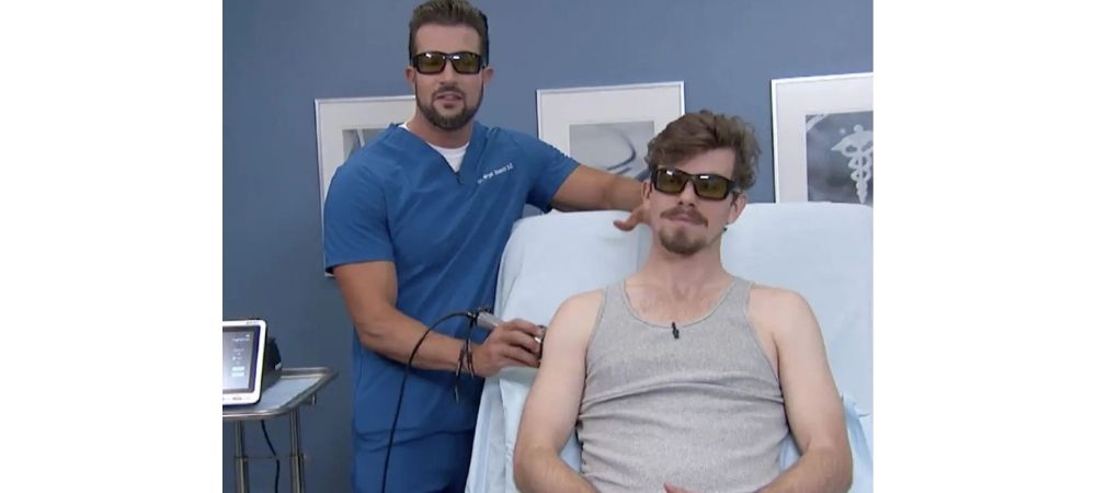
Can Laser Therapy for Pain Be a Viable Treatment?Learn what The Doctors have to say about laser therapy for pain. Watch Dr. Bryan Abasolo on the TV show, The Doctors as he treats patient Michael, a rock climber with shoulder pain, with a LightForce Therapy Laser and answers the question, ‘Can Deep Tissue Laser Therapy Treat Muscle Pain?’. Image Laser therapy is a safe and effective solution to relieving pain and healing the body without invasive surgery or potentially harmful medications. Yes, laser therapy for pain can be a viable option. Learn more about laser therapy.> Speaker One (00:02): Constant nagging Pain is frustrating and negatively affect your daily life could laser help believe it.Chiropractor Bryan Abasolois back in the procedure room with Michael, who is currently dealing with shoulder pain Michael, what’s going on with your shoulder, buddy? Michael (00:15): I wish I knew I’ve had a nagging shoulder injury for about a month now, and I’m an avid rock climber. I’ve been climbing two or three times a week for almost six years and haven’t been able to for the better part of a month, just because of this thing. And, you know, I’ve looked into different you know, ways of getting it healed without really an invasive under the knife procedure. So you know, I’m looking forward to seeing what this can do for me. Speaker One (00:38): So, Bryan, what do you think is going on? And what’s the game plan? Dr. Bryan Abasolo (00:41): Well, doc, what I think is going on, I think he has a little bit of what we call rotator cuff syndrome and specifically the superspinatus muscle is the actually most commonly injured muscle in the rotator cuff. So what we’re going to do today is a therapy called deep tissue laser therapy [for pain relief]. Okay. it’s an FDA cleared a modality to treat pain and inflammation. It’s going to help out tremendously with the recovery time of the injury. And what I love about it is that it’s safe. It’s painless, it’s noninvasive. And another great aspect of it is it’s very quick. Treatments are only about five to 10 minutes on average and believe it or not, this is actually used in NBA locker rooms, NFL locker rooms to get their athletes back on the field quicker. So I think Michael is going to do great with this modality. Speaker Two (01:32): So Bryan, we have lasers for everything. I’m fascinated here. What specifically, what kind of chiropractic laser is this? What wavelength obviously it’s meant to penetrate deeper into the tissues? Dr. Bryan Abasolo (01:46): Yeah, so this is not to be confused with a cold laser. That’s a level three laser. This is actually a step above. This is a class four laser it’s actually one of the strongest, the strongest therapeutic laser on the market. So what it’s going to do is the laser energy is actually going to penetrate deep down into the cells and the effect it’s going to have on the cells is actually going to penetrate the mitochondria, which is basically the powerhouse of the cell. And it’s going to produce more ATP, which is our energy currency. And when ATP is ramped up, cellular regeneration cellular metabolism is increased and that’s going to really help reduce pain, inflammation and increased range of motion. Speaker Two (02:28): Alright. Get started there, Bryan We all go through these sports injuries and so many different options. If you can comment how you would compare this laser treatment with the other things that we’re trying, we’re doing the PRP, we’re doing shock pulse therapy other forms of massage. Dr. Bryan Abasolo (02:52): Yeah. This is a great junk therapy you could use with a lot of things that doctors and PTs and chiros are doing now in physical therapy. You name it, electrical muscle stimulation, adjustments, you know, obviously some ultrasound you can do, but there’s nothing like this treatment at all. As far as the depth that it can penetrate deep down to the cells. Speaker Three (03:15): Michael, do you feel anything Michael (03:17): Feels like a nice massage at the moment. Dr. Bryan Abasolo (03:20): Yeah. So what I’m doing right now, he should start to feel a little, a nice soothing warmth and the laser actually has different treatment heads. So the one I’m using right now, it actually adds a massage component to it. So not only is he getting a nice therapeutic massage, but also the laser is penetrating into the cells of the rotator cuff, Dr. Bryan Abosolo (03:41): How much Bryan would something like this cost for one treatment and then in the case of someone like Michael, who has just this probably little nagging rotator cuff issue, how many treatments would you suggest? Dr. Bryan Abosolo (03:52): Yeah, I would say on average it can range anywhere from $50 to $80, depending on where you’re at in the country and the market. As far as how many treatments I would say for acute injuries, kind of like what Michael has right now, I would say a good number to really start feeling an effect is three to five visits with a magic number being at six and something more chronic like, you know, back pain that’s lasted for five years. I would say 10 visits is a good number to really start seeing some relief. Speaker One (04:21): Bryan, it seems like this is a great option for folks who have exactly what Michael has and Michael you’re anxious to get back out there and climb. Aren’t you bud? Michael (04:31): You have no idea. Speaker One (04:32): Good stuff. Well, best of luck to you, Michael and Bryan, thank you so much for sharing that device with us. Read More Blog Posts

Blog post
Cryotherapy vs. Laser Therapy for Treating InflammationContributed by Mark Callanen, PT, DPT, OCSDirector of Clinical Development, LightForce® Therapy Lasers Inflammation is a topic that is discussed in the clinical setting on a daily basis. It is an important part of the healing process and is needed for the proper healing of acute soft tissue injuries.1 Patients often are seeking relief from the associated pain and seek advice on how to best manage these injuries. How well a clinician understands the inflammatory process and how various anti-inflammatory modalities impact the healing process can have a profound effect on both the recovery time and the tissue repair process for damaged muscle tissue. Acute Inflammation (NSAIDS) There is rising evidence that dampening acute inflammation impairs muscle growth and regeneration in animal and human models.2 Despite this fact, clinicians are often quick to recommend NSAIDs to address acute inflammation due to their ability to decrease pain and reduce inflammation via their impact on cyclooxygenase-2 (COX-2) enzymes. While decreasing swelling, NSAIDs have been shown to negatively impact tissue repair by diminishing proliferation, differentiation, and fusion of satellite cells in muscle tissue which can lead to impaired skeletal muscle repair and growth, and increased fibrosis.3,4 Accordingly, it is not surprising that recent studies suggest many therapists have incomplete or inadequate knowledge on the topic of NSAIDS5,6 or have failed to upgrade their knowledge on the topic.7 This has the potential to be a significant problem in the management of musculoskeletal injuries as physical therapists are assuming increased roles in the direct access market. It is important for clinicians to understand that COX-2 enzymes are involved with both pro-inflammatory and anti-inflammatory mediators at different stages of the inflammatory process.8,9 While blocking COX-2 activity can reduce a pro-inflammatory response it can also inhibit the resolution of inflammation which can negatively impact muscle healing.2 Additionally, there are a variety of inflammatory mediators that play important messenger roles regarding the recruitment of neutrophils and monocytes that are essential for tissue repair. Inhibiting the inflammatory process can impair macrophage collection at the injured site which negatively impacts tissue repair.2 This can include persistent necrotic myofibers, increased fat accumulation10, decreased myofibril size, and slowed muscle regeneration.11 Consideration should be given to these factors before recommending NSAIDS for acute muscle injuries. Chronic Inflammation (NSAIDS) When advising patients on the topic of chronic inflammation, clinicians should be aware that NSAIDS have been found to be ineffective in reducing diffuse, systemic inflammation.12 Steroids (SAIDs) such as prednisone have been found to be a better alternative in restoring the balance of the inflammatory process for chronic inflammation2, especially with repeat “asynchronous” injuries to muscle tissue, such as repeated muscle strain to the same tissue.11,13 Controlling chronic inflammation is important as it can increase muscle degradation and reduce the synthesis of muscle fiber contractile proteins.14 Examples of chronic inflammation include diffuse OA, chronic COPD, aging, and kidney disease, all of which are associated with muscle wasting.15,16 It should be noted the positive effects of SAIDS is limited to the window: months to years.17,18 Prolonged use can lead to protein degradation which leads to muscle atrophy as well as decreased proliferation and differentiation of myoblasts.19 Long term muscle wasting outweighs short term anti-inflammatory benefits in many cases. Take Home for Anti-Inflammatory Medications Local inflammation is needed and benefits the healing process. Analgesics and analgesic modalities are a better choice than NSAIDS for acute muscle injuries. Chronic local conditions, NSAIDS help via pain relief, but are not good at reducing chronic inflammation. SAID use is warranted for chronic, local inflammation, however; prolonged use can lead to protein breakdown and promote muscle wasting. They should be used judiciously. Ice/Cryotherapy What about ice, sometimes referred to as cryotherapy? People have been using it for decades to manage inflammation so it must be a good choice, or is it? In 1978 an orthopedic surgeon, Gabe Mirkin, M.D. came up with the familiar acronym “R.I.C.E.” (Rest, Ice, Compression, and Elevation) regarding how to address inflammation. It’s simplicity and general adoption has been the gold standard for addressing inflamed tissue in the medical community for almost 40 years. The tide currently appears to be changing. Dr. Mirkin himself was recently quoted in an article published on the Spartan website (www.life.spartan.com) that he now openly rejects at least half of R.I.C.E. “I do not believe in cooling anymore,” he explained via email. Nor does he believe in the “R” component of his famous prescription either. In a foreword to the second edition of Iced!, Dr. Mirkin says most athletes are far more concerned with long-term healing than transient pain relief. “And research,” he writes, “now shows that both ice <or cryotherapy> and prolonged rest. actually delay recovery.” The research he may be referring to includes a study in the British Journal of Sports Medicine that retrospectively investigated 22 separate studies and concluded that “ice is commonly used after acute muscle strains, but there are no clinical studies of its effectiveness.” More damning was the report in the Journal of Strength and the Conditioning Research which stated that not only does cryotherapy fail to help injuries heal, it may well delay recovery from injury.20 Ice apparently plays more of an analgesic role post injury more than it does in reducing inflammation. A 2013 study in the Journal of Applied Physiology showed that cryotherapy had null to mild impact on pro-inflammatory markers on post-exercise induced muscle damage.21 Research from the Cleveland Clinic has gone a step further showing that Icing an injury delays the release of IGF-1 (insulin-like growth factor-1), a hormone involved in the inflammatory cascade that helps repair damaged tissues.22 These findings should at least start to bring into question the effectiveness of cryotherapy. Laser So if R.I.C.E. is no longer the answer, and NSAIDS are detrimental to the tissue healing process for acute injuries and ineffective for chronic inflammation, what’s a better strategy for managing inflammation and expediting tissue repair? Promoting active recovery is becoming much more accepted as the preferred plan of care. Gary Reinl, veteran athletic trainer and author of the book Iced! The Illusionary Treatment Option believes the answer lies in a new acronym: A.R.I.T.A.—Active Recovery Is The Answer. Instead of being still and shutting down blood blow, try to get things moving and circulating as soon as possible. In line with this concept, laser therapy is starting to gain traction in more forward-thinking training rooms and rehabilitation centers that have acquired this technology. The laser is instrumental in creating photobiomodulation (PBM) which is the mechanism by which photons elicit photophysical and photochemical events in tissues leading to physiological changes and therapeutic effects. This process helps hasten the inflammatory process which leads to improved tissue healing. Unlike NSAIDS which block the inflammatory cascade at the COX-2 level, and cryotherapy which delays the inflammatory process by restricting blood flow for a period of time, laser metabolically influences the injured tissue at the mitochondrial level, accelerating the healing process. PBM has both a direct photochemical influence on the mitochondria via Cytochrome C Oxidase23,24 and indirect modulation on the inflammatory cascade via enzymatic changes.25 Both of these effects decrease the length of time needed for tissue repair. Laser research that investigates the mechanisms involved with reducing inflammation, at a glance, look similar to pharmacological studies because they impact the inflammatory cascade at similar points. These include reduced COX-2 levels26, reduced Bradykinin levels27, reduction in IL-1 levels28, and reduction in PGE-2.29 It is important to understand that these reductions are fundamentally different with PBM in that they take place from intrinsic, anti-inflammatory signaling generated by better cell metabolism and improved micro-circulation. There have been two recent studies that have compared: ice, ice combined with laser, and laser therapy used independently to treat quadriceps muscles after maximum volitional contractions (MVC). They concluded that the laser had significantly higher MVC on retest and less oxidative stress compared to the placebo group. They also found that when cryotherapy was combined with the laser it lowered the efficacy of laser treatment done independently.30 A second study looked at similar groupings but looked at delayed onset muscle soreness (DOMS), MVC, and oxidative damage. They found that laser used as a single treatment (not performed in conjunction with ice) is “the best modality for enhancement of post-exercise restitution, leading to complete recovery to baseline levels from 24 h after high-intensity eccentric contractions.”31 These findings generally support the idea of active recovery. Specifically, they support the use of PBM to help with muscle recovery after exercise. These studies in conjunction with other emerging research are clearly bringing the use of cryotherapy and NSAIDS into question as the ideal choices for managing injured muscle tissue. It is never easy to change the way clinicians practice, but research is starting to suggest that the ideal way to address pain and inflammation will require a departure from past norms. Avoiding ice and NSAIDs in the early stages of the inflammatory process and introducing pro-metabolic modalities like laser therapy could become the new standard for evidence based practice. Clinicians might need to start wrapping their heads around the A.R.I.T.A. philosophy and put R.I.C.E. to REST. References Smith C, Kruger MJ, Smith RM, Myburgh KH. The inflammatory response to skeletal muscle injury: illuminating complexities. Sports Med . 2008;38:947–969. Duchesne E, Dufresne S, Dumont N. Impact of Inflammation and Anti-inflammatory Modalities on Skeletal Muscle Healing: From Fundamental Research to the Clinic. Physical Therapy [serial online]. August 2017;97(8):807-817. Bondesen BA, Mills ST, Pavlath GK. The COX-2 pathway regulates growth of atrophied muscle via multiple mechanisms. Am J Physiol Cell Physiol . 2006;290:C1651C1659. Bondesen BA, Mills ST, Kegley KM, Pavlath GK. The COX-2 pathway is essential during early stages of skeletal muscle regeneration. Am J Physiol Cell Physiol . 2004;287:C475–C483. Braund R, Abbott JH. Recommending NSAIDs and paracetamol: a survey of New Zealand physiotherapists’ knowledge and behaviours. Physiother Res Int . 2011;16:43–49. Green M, Norman KE. Knowledge and use of, and attitudes toward, non-steroidal anti-inflammatory drugs (NSAIDs) in practice: a survey of Ontario physiotherapists. Physiother Can . 2016;68:230–241. Grimmer K, Kumar S, Gilbert A, Milanese S. Non-steroidal anti-inflammatory drugs (NSAIDs): physiotherapists’ use, knowledge and attitudes. Aust J Physiother . 2002;48:82–92. Duchesne E, Tremblay MH, Côté CH. Mast cell tryptase stimulates myoblast proliferation; a mechanism relying on protease-activated receptor-2 and cyclooxygenase-2. BMC Musculoskelet Disord . 2011;12:235. Gilroy DW, Colville-Nash PR, Willis D et al Inducible cyclooxygenase may have anti-inflammatory properties. Nat Med . 1999;5:698–701. Summan M, Warren GL, Mercer RR et al Macrophages and skeletal muscle regeneration: a clodronate-containing liposome depletion study. Am J Physiol Regul Integr Comp Physiol . 2006;290:R1488–R1495. Arnold L, Henry A, Poron F et al Inflammatory monocytes recruited after skeletal muscle injury switch into anti-inflammatory macrophages to support myogenesis. J Exp Med . 2007;204:1057–1069. Sin DD, Reid WD. Is inflammation good, bad or irrelevant for skeletal muscles in COPD? Thorax . 2007;63:95–96. Dadgar S, Wang Z, Johnston H et al Asynchronous remodeling is a driver of failed regeneration in Duchenne muscular dystrophy. J Cell Biol . 2014;207:139–158. Bonaldo P, Sandri M. Cellular and molecular mechanisms of muscle atrophy. Dis Model Mech . 2013;6:25–39. Wåhlin-Larsson B, Carnac G, Kadi F. The influence of systemic inflammation on skeletal muscle in physically active elderly women. Age (Dordr) . 2014;36:9718. Batsis JA, Mackenzie TA, Jones JD et al Sarcopenia, sarcopenic obesity and inflammation: results from the 1999–2004 National Health and Nutrition Examination Survey. Clin Nutr. 2016;35:1472–1483. Sali A, Guerron AD, Gordish-Dressman H et al Glucocorticoid- treated mice are an inappropriate positive control for long-term preclinical studies in the mdx mouse. PLoS One . 2012;7:e34204. Manzur AY, Kuntzer T, Pike M, Swan A. Glucocorticoid corticosteroids for Duchenne muscular dystrophy. Cochrane Database Syst Rev . 2008;(1):CD003725. te Pas MF, de Jong PR, Verburg FJ. Glucocorticoid inhibition of C2C12 proliferation rate and differentiation capacity in relation to mRNA levels of the MRF gene family. Mol Biol Rep . 2000;27:87–98. Reinl G. ICED! The Illusionary Treatment Option. Oct 15, 2013: 9-11. Crystal NJ, Townson DH, Cook SB, LaRoche DP. Effect of cryotherapy on muscle recovery and inflammation following a bout of damaging exercise. Eur J Appl Physiol. 2013;113:2577–2586. H. Lu, D. Huang, N. Saederup, I. F. Charo, R. M. Ransohoff, Zhou. Macrophages recruited via CCR2 produce insulin-like growth factor-1 to repair acute skeletal muscle injury. The FASEB Journal, 2010; DOI: 10.1096/fj.10-171579 Chow R. et al. Inhibitory Effects of Laser Irradiation on Peripheral Mammalian Nerves and Relevance to Analgesic Effects: A Systematic Review. Photomedicine and Laser Surgery Volume X, Number X, 2011. Mary Ann Liebert, Inc. Pp. 1–17. Karu T et al. (1997) He-Ne laser radiation influences single-channel inonic currents through cell membranes: a patch-clamp study. Proc. SPIE. 3198:57-66. Liebert, A.D. et al. (2014) Protein conformational modulation by photons: a mechanism for laser treatment effects. Med Hypothesis. 82(3):275-281. Prianti, A.C.G. et al. (2014) Low-level PBMT (LLLT) reduces the COX-2 mRNA expression in both subplantar and total brain tissues in the model of peripheral inflammation induced by administration of carrageenan. Lasers Med Sci. 29(4):1397-1403. Jimbo, K. et al. (1998) Suppressive effects of low-power laser irradiation on bradykinin evoked action potentials in cultured murine dorsal root ganglion cells. Neurosci Lett. 240(2):93-96. Lopes-Martins, R.A. et al. (2005)Spontaneious effects of low-level PBMT (650 nm) in acute inflammatory mouse pleurisy induced by carrageenan. Photomed Laser Surg. 23(4):377-381. Mizutani, K. et al. (2004) A clinical study on serum prostaglandin E2 with low-level PBMT. Photomed Laser Surg. 22(6)537-539. De Marchi, T. et al. (2017) Does photobiomodulation therapy is better than cryotherapy in muscle recovery after a high-intensity exercise? A randomized, double-blind, placebo-controlled clinical trial. Lasers Med Sci. DOI 10.1007/s10103-016-2139-9. De Paiva, P. et al. Photobiomodulation therapy (PBMT) and/or cryotherapy in skeletal muscle restitution, what is better? A randomized, double-blinded, placebo-controlled clinical trial. Lasers Med Sci (2016) 31:1925–1933. Read More Blog Posts
Blog post
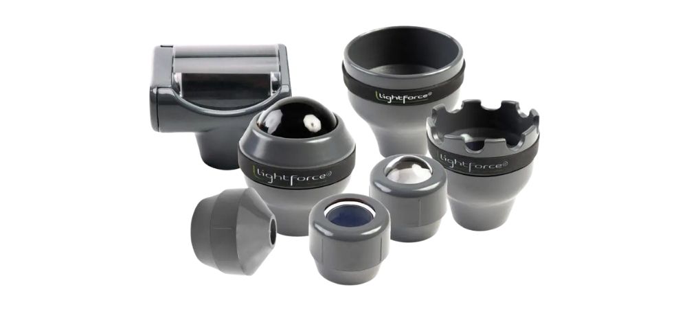
LightForce Laser Treatment HeadsWhat are the applications for different laser treatment heads? The following Exclusive LightForce Treatment Heads are available on all LightForce Therapy Lasers i-Series devices through the Empower and Empower IQ Delivery Systems. Image Small Cone Systems: 25W XPi, 15W FXi When treating very narrow or precise areas the Small Cone is ideal due to its smaller spot size. Small spaces can be treated easily by moving the smaller beam back and forth in areas that are difficult to treat with our larger heads. Recommended for use on fingers and when treating conditions on the face where you want to avoid light reaching the eye. Designed to be used with low to moderate power. Image Small Massage Ball Systems: 40W XLi, 25W XPi, 15W FXi When on contact treatment is needed in tight spaces, the Small Massage Ball is often the choice clinicians go to. Designed to be used with low to moderate power, it is ideal for treating on-contact around the face or around the digits of the feet and hands. Great for use over trigger points due to its ability to provide warmth and pressure to sore areas. Image Flat Window Systems: 40W XLi, 25W XPi, 15W FXi Our original treatment head has tons of versatility. Designed to be used for off-contact treatments around most areas of the body. Great for treating superficial tissues with small to moderate surface areas, especially in the presence of boney prominences. Due to its compact spot size it is also an option when treating cervical pathology that requires treating around the hair line. Should be used with low to moderate power settings. Image Large Massage Ball Systems: 40W XLi, 25W XPi, 15W FXi The patented Massage Ball is our most commonly used on-contact head. It helps clinicians achieve consistent results when treating deeper tissues due to its ability to maximize the delivery of light from your laser. The Massage Ball reduces light scatter on the surface while allowing physical compression of tissue. Paired with higher power settings, it is the ideal tool to address problems in the back, hips, and larger muscle groups. While effective, patients also love it due to its ability to couple soothing warmth with compressive massage over sore areas. This head can be used for all power settings and is ideal for treating deeper tissues with high power. Image Large Cone Systems: 40W XLi, 25W XPi, 15W FXi The Large Cone is designed for off-contact treatments that require higher power settings. Super versatile, it should be the go to head selection for most conditions where off contact treatment is desired up to 25 Watts. Ideal for treating larger areas or when deeper tissues need to be addressed. Not recommended for treatment around the face or over smaller areas due to its larger spot size. Image XL Cone System: 40W XLi The XL Cone was created for our 40 Watt XLi to allow users to confidently treat multiple conditions with our highest powered platform. Coupled with our patented Smart Hand Piece, the XL Cone has the unique ability to keep high power treatment comfortable for most patients. With a spot size that is twice the size of our Large Cone, it allows clinicians to safely treat large areas and deeper conditions at over 25 Watts of power. Ideal for use over the trunk, hips, and large extremities, it can be used with any power setting, but shines above 25 Watts. Image Rolling Pin System: 40W XLi LightForce created the Rolling Pin to allow large areas to be treated with the new 40W XLi with a one of a kind, on-contact head that makes treatments simple. Coupled with the Smart Hand Piece, the Rolling Pin applies a wide elliptical spot to deep tissues that allows large, flat areas of tissue to be covered in a few passes of the handpiece. It is analogous to painting a wall with a roller vs. a brush. Ideal when treating the back, hip, and long extremities with higher power. Learn more about how laser therapy works. > Read More Blog Posts
Blog post
Reflection on “Actively” Rehabbing after Medial Meniscectomy Contributed by Mark Callanen, PT, DPT, OCSDirector of Clinical Development, LightForce® Therapy Lasers Being an orthopedic physical therapist for over 20 years allows you to see a lot of post-operative knees. While in practice I tried to balance evidence-based approaches with acquired clinical knowledge to generate successful outcomes on a regular basis. Most would agree, this usually requires a combination of appropriate manual techniques, exercise-based treatments, a touch of psychology, and the use of a few choice modalities along the way. Recently I had a unique opportunity to experience the rehabilitation process from a patient’s perspective after having 50% of my right medial meniscus removed. I had been having significant clicking and medial joint line pain for 6 months prior to surgery. No spectacular episode to reflect back on other than years of wear and tear. Prior to surgery my knee was progressively worsening. The primary complaint was notable clicking as the knee approached terminal extension during hill walking and light treadmill jogging. Deep knee flexion was also uncomfortable and any quick lateral motion was not well tolerated. Once I accepted the fact that my almost 50-year-old cartilage needed some surgical help, I scheduled the orthopedic consult. After surgery, I decided I would play the part of both patient and therapist during my rehab. I was curious to see if my personal concepts regarding the management of acute inflammation would hold up. The surgery was scheduled on a Friday morning and I was home by lunch time. A text book posterior horn medial meniscectomy was performed without complication. The only other tissue that was addressed during the arthroscopy was the removal of thickened plica from my medial joint capsule. The knee was flushed with anesthetic (20 cc of 0.5% ropivacaine) after the procedure to help control pain. Image Pre-Op Image Post-Op Post-operative instructions were to keep the dressing in place for 5 days with instructions to keep the bandage dry while icing and elevating the knee as needed for up to 2 weeks. Instruction was also given to take 800 mg ibuprofen every 4-6 hours during that time and take opioids (Norco) for pain as needed. Regarding rehab, instructions were to start bending knee as tolerated and begin rehab at an out-patient PT facility the following week. Pretty standard instructions that most clinicians could predict in their sleep. I of course did almost NONE of these things. I removed the outer dressing within 2 hours of getting home to expose the skin and allow me to inspect the swelling. I covered the portal sites with band aids to prevent infection. Removing the bandages made it easier to bend the knee and range the joint. Exposing the skin also enabled laser treatments to begin on day one. 4-5 hours post-surgery, I had minimal pain due to the anesthetic that was injected in the knee. I wanted to take advantage of this time period with reduced pain to see how the knee was moving and if the quad was firing. To my surprise, I had about 80% of knee flexion ROM and the quad was still able to volitionally contract. Since I had quadriceps control, I was able to walk and bear weight almost immediately without significant pain or difficulty. I did not require an assistive device. My primary goals were to reduce swelling quickly in order to maintain volitional VMO control, and restore normal ROM as quickly as possible to avoid stiffness/scar formation in the capsule. Image Day 2 Post-Op Image Day 3 Post-Op So what tools did I plan to use to help accomplish these goals? Fortunately, as the Director of Clinical Development at LightForce Therapy Lasers, I have access to a Class IV laser and other devices that enabled me to quickly progress my program. In addition to the laser, I was also able to utilize 2 other modalities to assist during my recovery; an M-Trigger biofeedback unit as well as a negative pressure device (LymphaTouch) designed to help with swelling and scar tissue mobility. Given the current research on cryotherapy, I was not planning to utilize ice unless there was a significant amount of joint effusion.1 Luckily, that wasn’t the case. I focused on pursuing active healing in order to promote better blood flow and try to abate the inflammatory cascade. 4 hours after surgery I began lasering my knee to promote photobiomodulation. Treatment areas included all exposed areas of the joint and the distal quadriceps except for the incision sites. Treatment was performed in a circumferential fashion. Settings were in the 6-8 J/cm2 range for the first week. Power ranged from 6 Watts to start and progressed it by 2 Watts a day for the first two weeks. I capped power at 25 Watts. Treatment was performed 2 x day for the first 5 days, and every other day the second week due to the progress that was observed. Swelling improved each day due to the effects of photobiomodulation and volitional quadriceps activities. Open and closed chain strengthening started 2 days after surgery at a local gym. ROM was restored to WNL within 3 days after the surgery with pain never going above 3/10 at any point in the process. NSAIDS were discontinued after 2 days and the opioid prescription was never filled. I received the M-trigger a couple days after the surgery date. It quickly pointed out that my EMG activity was at about 50% of my non-involved quad when training it with quad sets and straight leg raises. This was not surprising, but at 2 weeks after surgery, where I had been doing unilateral leg extension exercises in a gym with 20-30 lbs. I was amazed to see that motor firing was still operating at a 20% deficit! I would have had no way to appreciate this without the M-Trigger device. A 2010 study confirmed that EMG activity is often decreased at 6 months after medial meniscectomy surgery. This deficit is present in cases where there is no difference in muscle control at submaximal levels and there is equivalent muscle architecture to the uninvolved side. The study concluded the deficit is likely attributable to neural impairments that affect muscle control at maximal force outputs.2 The study confirmed what I experienced. Even with minimal swelling at the knee, no significant pain, and a volitional muscle contraction in place while under a submaximal load, my medial quadriceps was not firing normally. It highlighted that retraining quadriceps activity after surgery is a longer process than I had previously appreciated. Not doing so increases the chance of reinjury for weekend warriors that return to sport with the quad in this state. Using tools like NMES and biofeedback should help address this problem. The last modality I was able to introduce to my program was a negative pressure LymphaTouch device. I was not able to apply it to my knee during my first week of rehab due to logistical reasons, which was unfortunate as it would have been great in helping with lymph flow around the knee. Since there was plica removed from my medial capsule, there was more swelling at the medial capsule than other parts of the knee. There were a handful of other areas that stood out as being sore with palpation: namely the bursa directly over the patella, the fibular head, and the lateral joint line proximal to the fibular head. Applying the LymphaTouch device with moderate suction, 125-175 mm Hg in an intermittent setting, helped with reducing residual swelling around the medial knee and patellar area during the 4 weeks after surgery. Using the device in a constant setting with higher suction (250 mm HG) allowed for myofascial treatments over areas that were more tender. The pulling and twisting of the tissue helped with soreness in the soft tissues that were difficult to address otherwise. Self-massage did not seem to impact it effectively. Treatment with the LymphaTouch was performed over the problem areas 3-4 times a week. This helped minimize soft tissue complaints by the end of week 4, where I was able to start kneeling on the knee with minimal pain. Once the incisions were completely closed at 3 weeks, the negative pressure treatments also helped reduce pain and swelling around the portal sites. Currently there is no abnormal tissue texture noted over the portal sites or any other area of the knee. Image 13 Days Post-Op Image 19 Days Post-Op Regarding exercise, a progressive lower extremity strength, balance, and ROM program started the day after surgery at a local gym. Activities on the day of surgery focused on heel slides, quad sets, and restoring normal gait mechanics. Starting on day 2, leg exercises were performed 3 x week in some variation of closed/open chain activity with a treadmill progression to return to jogging. Closed chain exercises were able to be introduced 2 days after surgery due to lack of pain and swelling. What did this active approach to knee rehab accomplish? Weight bearing started within hours of surgery. Over 90% of ROM was restored in 2 days and full ROM in 4 days. Volitional VMO control was never lost enabling hip and quad exercises to begin in the gym 2 days after surgery. Jogging in small one-minute bouts began in the second week. Prior to surgery I was unable to jog on a treadmill without pain/ clicking. NSAIDS were used for a 2 days after surgery, but all medicines were discontinued 3 days after surgery without every having pain over 3/10 level. Surgery was performed on a Friday and I was able to walk up to a second story the following Tuesday and sit comfortably at my desk for 6 hours. A couple closing notes. My surgery was performed by an experienced orthopedic physician. His ability to perform the procedure without unwanted disruption in other areas of the knee was evident by the lack of swelling after the treatment. A quick recovery is often dependent on a successful surgery. Another component that may have impacted post-operative swelling was a phenomenon referred to as preconditioning.2 My knee was treated with high power laser 2-3x week for several months prior to surgery in order to control swelling and pain. I walked into surgery without a pain complaint. Preconditioning has been shown to help reduce inflammatory responses by preventing apoptosis when cells are placed under oxidative stress. It may have been another component as to why I experienced limited swelling after the surgery. In a perfect world I would have started the negative pressure treatments on day one to assist the lymph system, but introducing this technology a week after surgery did not seem to negatively impact my progress. Starting the assault on the inflammatory cascade early on with the laser seemed to play a huge part in my painless progression into moderate level activity in less than a week. Rehabilitation specialists should also be acutely aware of the neural deficits that may be lingering during and after the subacute stage of recovery. Progressing patients to higher level activities based solely on observational findings could prove to be detrimental to the knee if muscle function is not assessed with some form of biofeedback. Finally, for those clinicians that are still heavily invested in cold modalities; it is worth reiterating that going with an active rehabilitation program is what drove this success story. Cryotherapy was not used at any point following surgery to address pain or inflammation. References Reinl G. ICED! The Illusionary Treatment Option. Oct 15, 2013: 9-11. Glatthorn JF, Berendts AM, Bizzini M, Munzinger U, Maffiuletti NA. Neuromuscular function after arthroscopic partial meniscectomy. Clin Orthop Relat Res. 2010;468(5):1336-1343. doi:10.1007/s11999-009-1172-4 Agrawal T, Gupta GK, Rai V, Carroll JD, Hamblin MR. Pre-Conditioning with Low-Level Laser (Light) Therapy: Light before the Storm. Dose-Response. October 2014. Read More Blog Posts
Blog post
Ever Wish You Had a Magic Wand to Help Treat Injured Tissue?Explaining how therapeutic laser can accelerate a plan of care. Contributed by Mark Callanen, PT, DPT, OCSDirector of Clinical Development, LightForce® Therapy Lasers Progressing plans of care (POC) is a careful dance between identifying the current stage of healing, balancing subjective pain complaints, and appropriately increasing therapeutic activity levels. Incorporating high power laser to create temporary analgesic effects and spur on tissue healing via photobiomodulation (PBM) may make this task easier for clinicians. Treating acute injuries usually focuses on controlling symptoms associated with pain and inflammation; which can include multiple techniques and modalities that will ultimately allow ROM and function to be restored later in the POC. This can be a trying time for athletes as their overall activity levels are reduced while their injury heals. What If? What if you could wave a magic wand over the injured area to reduce pain complaints (in minutes) and promote better ROM earlier in the POC? High power therapeutic lasers may be a modality that helps make this wish become a reality. Laser therapy promotes photobiomodulation (PBM) which is the mechanism by which photons elicit photophysical and photochemical events in tissues leading to physiological changes and therapeutic effects.1 Recent PBM research has shown that treating afferent nerves with higher power densities (irradiance) significantly impacts pain perception.2,3 When laser is applied in higher doses, it can slow down conduction rates and decrease the size of compound action potentials in both C and A delta afferent nerves.2,3 This can result in quick changes in patients’ pain complaints via true analgesia. The Results While getting quick pain relief is a desirable benefit when using high power laser, most clinicians want to achieve lasting results. The good news is that you don’t have to sacrifice one for the other. High power laser has the potential to help with both short term pain blocking and improved long-term function. A 2018 study out of The University of Cairo looked at patients with rotator cuff tendinopathy that underwent exercise programs combined with either high intensity laser (HILT) or sham laser treatments, performed 3 x week for 4 weeks. The HILT group had significant improvements in pain, ROM, and Shoulder Pain and Disability Index (SPADI) scores compared to the sham group. Significant differences were still noted at 3 and 6 months.4 Laser can be used for a variety of diagnoses. In 2017 The American College of Physicians endorsed Low Level Laser Therapy (LLLT) as the only passive modality in its treatment recommendations for chronic low back pain.5 The Journal of Orthopedic and Sports Physical Therapy (JOSPT) similarly recommended laser in its 2014 Clinical Practice Guidelines for the treatment of heel pain and plantar fasciitis6 as well as its 2017 Revision of Neck Pain Treatment Guidelines, where it recommended laser in two subcategories.7 Consistent Outcomes Keys to getting consistent outcomes with any laser device is understanding how power, time, and wavelength impact dosing, while knowing the specifications of the device being used is imperative. With correct dosing and adequate irradiance, PBM should open the door in the early phases of a POC to progress exercise programs and provide treatment options that would otherwise be delayed due to pain. This factor combined with the fact that PBM has been shown to help improve tissue healing through all phases of tissue healing, could result in athletes magically getting back on the field ahead of schedule. Learn More > References 1. Anders JJ, Lanzafame RJ, Arany PR. Low-level light/laser therapy versus photobiomodulation therapy. Photomed Laser Surg 2015;33:183–184. 2. Chow et al. Inhibitory Effects of Laser Irradiation on Peripheral Mammalian Nerves and Relevance to Analgesic Effects: A Systematic Review. Photomedicine and Laser Surgery Volume X, Number X, 2011ª Mary Ann Liebert, Inc. Pp. 1–17. 3. Holanda, V.M. et al. (2017) The Mechanistic Basis for Photobiomodulation Therapy of Neuropathic Pain by Near Infrared Laser Light. Lasers Surg Med. 2017 Jul;49(5):516-524. 4. Elsodany, A. et al. Long-Term Effect of Pulsed Nd:YAG Laser in the Treatment of Patients with Rotator Cuff Tendinopathy: A Randomized Controlled Trial. Photomedicine and Laser Surgery, Vol 36 (9), 2018, pp. 506–513. 5. Qaseem A, Wilt TJ, McLean RM, et al, for the Clinical Guidelines Committee of the American College of Physicians. Noninvasive Treatments for Acute, Subacute, and Chronic Low Back Pain: A Clinical Practice Guideline From the American College of Physicians. Ann Intern Med. 2017;166:514–530. https://annals.org/aim/fullarticle/2603228/noninvasive-treatments-acute-subacute-chronic-low-back-pain-clinical-practice?_ga=2.94313501.1892251386.1574364066-1113658412.1572282428 6. Martin, R, et al. Heel Pain—Plantar Fasciitis: Revision 2014 Clinical Practice Guidelines Linked to the International Classification of Functioning, Disability and Health From the Orthopaedic Section of the American Physical Therapy Association. J Orthop Sports Phys Ther. 2014;44(11): A1-A23. 7. Blanpied, P, et al. Neck Pain: Revision 2017 Clinical Practice Guidelines Linked to the International Classification of Functioning, Disability and Health From the Orthopaedic Section of the American Physical Therapy Association. J Orthop Sports Phys Ther. 2017;47(7): A1-A83. Read More Blog Posts